Restriction Digest Not Cutting or Wrong Bands
7 min read · Updated July 10, 2026
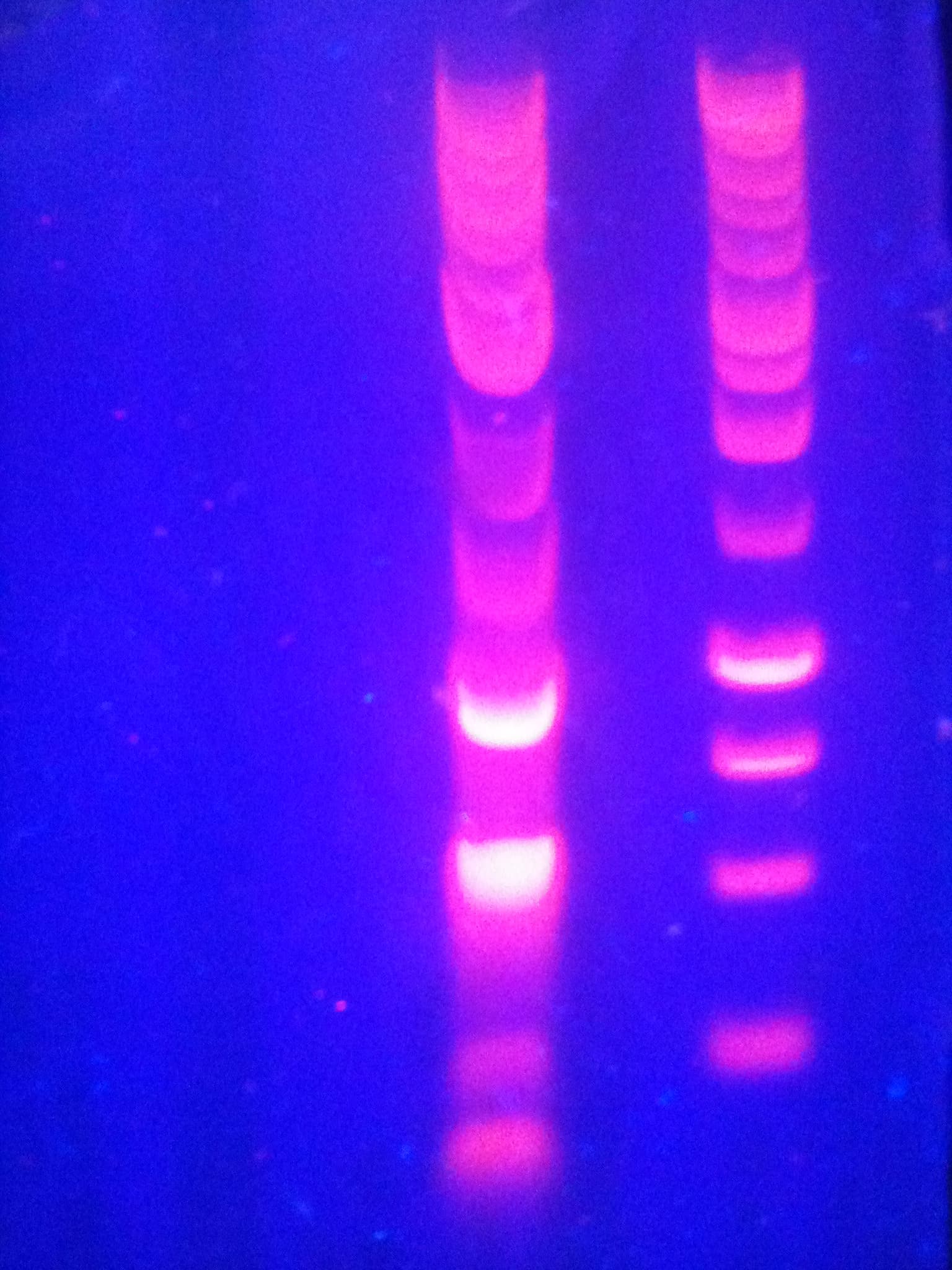
A restriction digest that comes back looking wrong — one uncut band, too few bands, extra bands, or an uncut control lane sitting at a size that does not match the plasmid map — is one of the most common places a cloning or diagnostic workflow stalls. Most of the time the cause is not a mystery: it falls into a short list of known failure modes, and the fastest way to identify which one you are looking at is to know what each looks like on a gel. This guide works through the digest problems molecular biologists run into most often, organized by symptom, with the likely cause and the fix for each.
No cutting at all: the digest lane looks identical to the uncut control
If the digest lane runs as a single band indistinguishable from undigested plasmid, the enzyme did not cut. The usual suspects are a short list, worth working through roughly in this order.
- Methylation-sensitive site in dam+/dcm+ prepped DNA. Standard cloning strains methylate DNA at Dam (GATC) and Dcm (CCWGG) sequences, and a number of restriction enzymes simply cannot cut when their recognition site overlaps a methylated base. If the plasmid came from an ordinary dam+/dcm+ strain and the enzyme is methylation-sensitive, it will not cut regardless of how good the buffer, temperature, or enzyme are — a commonly overlooked cause distinct from an enzyme or reaction failure.
- Wrong buffer or temperature for that specific enzyme. Each enzyme has a defined optimal buffer and incubation temperature, and running it outside those conditions can drop activity to near zero without any obvious sign in the reaction tube.
- Heat-inactivated, old, or improperly stored enzyme. Repeated freeze-thaw cycles or storage above the recommended temperature erode activity over time; a parallel digest on DNA already known to cut with that enzyme tells you whether the problem is the enzyme or the template.
- The site you expected is not actually present. Confirm the recognition site is really in your sequence with an actual scan rather than eyeballing it, especially for degenerate, IUPAC-coded sites such as HinfI's G^ANTC, where the middle position can be any base and is easy to misjudge by eye.
Fewer bands than expected, or bands at the wrong sizes
When the digest clearly did something but the pattern does not match what you predicted, the cause is usually one of the following.
- Partial or incomplete digestion. Extending the incubation time or adding more enzyme often fixes it; if neither does, suspect an inhibitor carried over from the miniprep — residual salt, ethanol, or phenol can suppress enzyme activity even when the DNA looks clean by absorbance.
- A topology mismatch between what you assumed and what the digest actually produced. A circular plasmid with N cut sites gives N fragments, because there are no free ends, while a linear template with the same N cut sites gives N+1 fragments, since the two ends of the molecule count as fragment boundaries too. Predicting counts or sizes for the wrong topology makes a perfectly good digest look wrong.
- The wrong construct, or a contaminated prep. It is worth confirming the digest was actually run on the plasmid you think it was, particularly after a colony pick or a prep handled alongside other constructs.
Extra or unexpected bands beyond what was predicted
Extra bands usually mean one of two things, and predicting the expected pattern first is the fastest way to tell them apart.
- Star activity: relaxed, near-cognate site cutting caused by non-standard reaction conditions, such as too much glycerol carried in with the enzyme, excess enzyme relative to DNA, extended incubation, or the wrong ionic strength or pH. It becomes more likely the further a reaction sits outside the enzyme's recommended conditions, and it typically adds faint extra bands on top of the expected pattern rather than replacing it outright.
- The template genuinely is not what you think it is. A wrong clone, a mixed colony, or cross-contamination between preps produces a pattern that does not resemble your construct at all, rather than a mostly correct pattern with a few faint extras.
An uncut control that runs at an unexpected size
An uncut plasmid lane that does not land where its length would suggest usually is not a digestion problem at all — it is a property of DNA topology. Supercoiled circular DNA folds into a compact shape that migrates faster through agarose than a linear molecule of the same length, so an intact plasmid typically looks smaller than its actual size. Nicked or relaxed circular DNA, by contrast, migrates more slowly and looks larger.
Only linearized fragments — the ones a completed digest actually produces — migrate at a size consistent with their true base-pair length. An uncut control lane sitting at an unexpected size next to the ladder is expected behavior, not evidence that something went wrong with the digest.
Predict the digest before you troubleshoot the gel
Every symptom above resolves faster once you know what the digest was supposed to produce. Working out the exact fragment count and sizes for the actual topology, linear or circular, before you run anything turns a vague sense that something is off into a specific, band-by-band comparison, and that comparison is what actually distinguishes partial digestion, star activity, and a mislabeled construct from one another.
Virtual Gel takes your sequence and a chosen enzyme, computes the resulting fragments for either topology, and renders the simulated agarose gel with a ladder, so you have a stated expectation before you touch a pipette. Restriction Sites confirms which recognition sites are actually present on your sequence and where, including degenerate IUPAC-coded sites that are easy to miscount by eye, settling whether the site is really there directly instead of by inspection. Plasmid Viewer places those same cut sites on a circular or linear map of your construct, useful for checking whether a digest was run on the clone you actually think it was.
Frequently asked questions
Why is my restriction digest not cutting at all?
Check whether the enzyme is methylation-sensitive and the DNA came from a standard dam+/dcm+ cloning strain, since that combination will not cut regardless of buffer or temperature. Also rule out the wrong buffer or incubation temperature, a heat-inactivated or old enzyme, and confirm the recognition site is actually present in your sequence rather than assumed.
Why does my restriction digest have fewer bands than expected?
The most common causes are partial digestion, which more enzyme or more time usually fixes, and a topology mismatch — a circular plasmid with N cut sites gives N fragments, while a linear template with the same N sites gives N+1 fragments. Confirm which topology you actually predicted for before assuming the reaction failed.
What causes extra bands in a restriction digest?
Extra bands usually mean star activity from non-standard buffer, salt, or glycerol conditions and enzyme overuse, or that the template is not what you think it is. Predicting the expected fragment pattern first and comparing it band for band against the real gel is the fastest way to tell which explanation applies.
Why does my uncut plasmid run at an unexpected size on a gel?
Supercoiled circular DNA migrates faster than its actual length and looks smaller, while nicked or relaxed circular DNA migrates slower and looks larger. Only linearized digest fragments migrate at a size consistent with their true base-pair length, so an odd-looking uncut lane is expected, not a sign of a failed digest.
Related references
Common restriction enzymes: recognition sites, cut positions, NEB buffer activity, star activity and an interactive double-digest buffer finder.
Nucleotide ambiguity codes and their complements.
Selection markers, mechanisms and working concentrations for cloning.