How to Verify a Clone with Sanger Sequencing
6 min read · Updated July 10, 2026
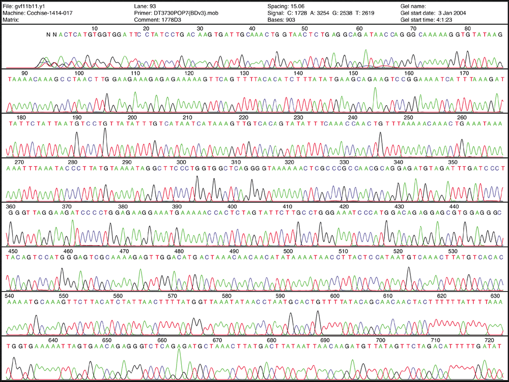
You cloned an insert, sent it for Sanger sequencing, and now have a chromatogram and a reference sequence you need to reconcile. The hard part is rarely finding the file — it's knowing which peaks to trust, whether a stacked double peak means anything, and whether a mismatch against your reference is a real mutation or just sequencing noise. This guide walks through reading the trace, trimming to the usable region, telling point mutations apart from indels, and aligning what's left to your expected sequence.
What a chromatogram is actually showing you
An .ab1/.abi file is not a sequence, it's four traces. At every base position the sequencer records signal from four dyes, and the software calls a base wherever one trace peaks well above the other three. Under the ABI convention you'll usually see green for A, red for T, black for G, and blue for C, plotted underneath the called-base letters. Each base also carries a quality value, similar in spirit to the Phred scores you'd see from short-read platforms, and that number is doing a lot of the work when you decide whether to believe a given position.
Not all of the read deserves your trust
A Sanger read of 700-900 usable bases is not uniformly reliable end to end. The first 20-35 bases are typically the worst part of the trace, and it is a resolution limit rather than a failed reaction. Applied Biosystems states that the properties of the dyes and the separation polymer together limit resolution over the first 20-25 bases, and that poor quality through the first 35 is normal with its standard chemistry. Quality climbs after that, holds through a long, well-separated middle stretch, and then degrades again well before the physical end of the read as peaks broaden and merge.
- Discard or heavily discount the first 20-35 bases regardless of how clean they look.
- Treat the tapering final stretch the same way — it isn't the true end of your insert, it's the point where the sequencer stopped resolving well.
- Do your actual comparison work on the wide, evenly spaced middle of the trace, which is where the base calls and quality scores are most trustworthy.
Double peaks versus messy peaks — two different problems
A single stacked or overlapping double peak at one position, with clean single peaks on either side, almost always means the template wasn't pure at that base — a heterozygous site, or a bacterial culture or PCR pool carrying both a wild-type and an edited sequence. It's localized: one position, then back to normal.
An insertion or deletion looks completely different. Because the sequencer calls bases by position along the capillary run, one extra or missing base throws every subsequent position out of register, so everything downstream of an indel turns into superimposed, unreadable peaks for the rest of the read. That cascading mess is the signature of a frameshift, not a point mutation, and it's usually obvious once you know to look for it — a single messy position is a mixed template, a read that goes messy and stays messy is an indel.
Aligning the trimmed read to your reference
Once you've trimmed the noisy ends, the remaining high-quality stretch is what you actually align against your expected sequence. Two things trip people up here. First, orientation: your read may have come off either the forward or reverse primer, so if a straightforward alignment looks like garbage, try the reverse complement before assuming the clone is wrong. Second, flanking sequence: reads from a plasmid often include a bit of vector backbone on either side of the insert, so don't expect the whole read to match your insert reference base for base — expect a clean match in the region that corresponds to your insert, with vector sequence bracketing it.
Deciding whether a mismatch is real
Once aligned, you'll almost never get a perfect match across the whole usable region, and that's expected. What matters is where the mismatch sits. A single mismatched base that falls inside the low-quality first 20-35 bases, in the degraded tail, or on a peak that was ambiguous or poorly separated to begin with is more likely a sequencing error than biology. A mismatch sitting in the middle of a clean, high-quality, unambiguous stretch of trace — well-separated peaks on both sides — is much more likely to be a genuine change in your clone and worth following up with a repeat read or a second primer.
Where SeqBench fits in
Doing this by eye, base by base, across a 700-900 base read is slow and easy to get wrong. Sanger vs Reference takes your Sanger read and your expected reference sequence, aligns them end-to-end, and returns a pass / needs-review verification report with every mismatch and indel, so you can jump straight to the positions that matter instead of scanning the whole trace manually. If you want to look at the raw peaks first — the colors, the quality track, the double peaks — open the .ab1/.abi file in the Sanger Trace Viewer. And if what you're really checking is a specific set of expected edits (a substitution, an inserted tag, a deletion) rather than a general comparison, the Variant Comparator will align query to reference and list out the substitutions, insertions and deletions with their effects.
Frequently asked questions
How do I read a Sanger sequencing chromatogram?
Each peak is one base call, colored by dye (commonly green=A, red=T, black=G, blue=C under the ABI convention), with a quality value under it similar in spirit to a Phred score. Trust the well-separated, evenly spaced peaks in the middle of the read more than the compressed ones at either end.
Why does my Sanger sequencing result have two peaks stacked at the same position?
A single stacked double peak at one position usually means you sequenced a mixed template, such as a bacterial culture or PCR pool containing both wild-type and edited sequence at that base. If every peak after that position also turns messy, that pattern points to an insertion or deletion instead, not a clean point mutation.
How do I verify a clone by Sanger sequencing?
Trim the low-quality first 20-35 bases and the degraded tail, then align the remaining high-quality stretch against your expected reference in both orientations, since the read can come off either strand or include flanking vector sequence. Any mismatches that fall in clean, high-quality trace are worth following up; ones near the noisy ends usually are not.
How much of the trace should I actually trust?
Not the whole read. Discount the noisy bases at the very start, where the trace has not yet resolved away from the primer, and the degraded tail, where peaks broaden and merge as resolution runs out. Judge a mismatch by the local trace quality around it: one sitting in a clean, well-separated stretch is worth following up, while one at either noisy end usually is not. If you cannot open the file at all, that is a different problem — see the guide on opening an .ab1 file.
I picked eight colonies and got a zip back. Do I do this eight times?
No, and doing it one trace at a time costs you the check that matters most. Judge the plate together instead: reads are grouped into clones from their file names, every clone's reads are piled up in reference coordinates, and a difference one read reports at a position other reads cover is then reported as the sequencing error it is rather than as a mutation. One read on its own cannot make that distinction, because there is nothing to disagree with it. A position no read covered is never passed either, which is the other thing a per-trace pass silently gets wrong on a 6 kb plasmid read by one 900 bp primer.
Related references
Related tools
Inspect chromatograms, compare a read with its reference, or screen a whole plate of clones.
Inspect chromatograms, compare a read with its reference, or screen a whole plate of clones.
Paste what you got back and the PCR you say made the insert. It re-derives the insert, finds it in either orientation, and gives you the exact mismatch positions.
Align a query to a reference and list substitutions, insertions and deletions with effects.
Align your reads to your own annotated reference and get what each difference DOES — silent, missense at a named codon, or a premature STOP that ends the protein — not just where it sits.